
Junqi Liang 1, Zijun Zhang 2, Ahyan Ilman Qudsi 2, Qingfeng Liang 2
1 University College London Hospital, London, United Kingdom
2 Beijing Institute of Ophthalmology, Beijing Tongren Eye Center, Beijing Tongren Hospital, Capital Medical University, Beijing Key Laboratory of Ophthalmology and Visual Sciences, Beijing, 100005, China
Summary
Purpose To report fulminant Pseudomonas aeruginosa keratitis after bilateral ptosis correction in a child with Noonan syndrome, managed medically despite severe stromal thinning. A 12-year-old girl with genetically confirmed Noonan syndrome presented five days after bilateral ptosis correction with a red, swollen right eye and two days of visual loss unresponsive to topical tobramycin. Visual acuity was counting fingers in the right eye and 0.8 (decimal) in the left. Both upper lids were oedematous with lagophthalmos, and the right eye showed chemosis, lower-lid ectropion and bulbar conjunctival exposure. A 3 × 5 mm superior ulcer with dense infiltrate reached the pupillary zone; there was no hypopyon. Scrapings showed neutrophils and Gram-negative bacilli, and in vivo confocal microscopy showed inflammatory infiltration without hyphae or cysts. Culture grew P. aeruginosa. Fortified ceftazidime 50 mg/mL and levofloxacin 0.5% were alternated every 15 minutes for 24 hours, with cycloplegia, lubrication and intravenous ceftazidime. The infiltrate regressed by day 5 while the ulcer bed thinned markedly; low-concentration topical corticosteroid was then added under antibiotic cover. Keratoplasty was considered on day 10 and deferred in favour of continued medical therapy, intensive surface protection and bandage contact lens. By day 21 the cornea was intact and acuity had returned to 0.8. Conclusions Ptosis surgery may further compromise corneal protection in syndromic children. Persistent thinning after microbial control may reflect inflammatory collagenolysis rather than ongoing infection and does not necessarily require keratoplasty. If the infiltrate is resolving, the globe remains intact, and exposure can be corrected, the native cornea may be preserved.
Keywords: Pseudomonas aeruginosa; bacterial keratitis; ptosis surgery; exposure keratopathy; corneal melting
Introduction
Noonan syndrome is a genetically heterogeneous disorder caused by dysregulation of the RAS/MAPK signaling pathway. It is characterized by distinctive facial features, short stature, skeletal abnormalities, congenital heart disease, and variable developmental abnormalities(1). Ocular involvement is very common, affecting nearly all patients. In the prospective series of van Trier and colleagues, ptosis was present in 56% and lagophthalmos in 28%, alongside hypertelorism, epicanthal folds, refractive error, strabismus, and a range of anterior and posterior segment findings (2–3). The eyelid phenotype is clinically important because it directly affects corneal protection.
Ptosis correction may further compromise eyelid closure, leading to postoperative lagophthalmos and exposure keratopathy after either levator-based procedures or frontalis suspension (4–7). When persistent, corneal exposure can disrupt the epithelial barrier and create a vulnerable ocular surface that is more susceptible to secondary infection. In this setting, Pseudomonas aeruginosa is particularly concerning because it can cause rapid corneal destruction. Bacterial proteases directly degrade the stromal matrix, while the accompanying neutrophilic inflammatory response further accelerates collagen breakdown. Thus, an initially limited epithelial defect may rapidly progress to stromal melting, severe thinning, or impending perforation within days (8). Prompt recognition of infection, rapid identification of the causative pathogen, and intensive antimicrobial therapy are therefore essential to prevent irreversible corneal damage and vision loss (9).
We describe a child with Noonan syndrome who developed fulminant P. aeruginosa keratitis shortly after bilateral ptosis correction. Despite the rapid progression of infection, the cornea was successfully preserved without transplantation.
Case Report
A 12-year-old girl was referred to our tertiary eye center five days after undergoing bilateral ptosis correction at another hospital. She presented with redness and swelling of the right eye and a two-day history of severe visual loss. Topical tobramycin prescribed at the referring hospital had not improved her symptoms.
She had a history of growth and developmental delay since early childhood, together with a chest-wall deformity and congenital heart disease. Noonan syndrome had previously been confirmed by molecular genetic testing at another institution. She had no history of ocular trauma, contact lens wear, or ocular surgery apart from the recent ptosis procedure.
On presentation, visual acuity was counting fingers in the right eye and 0.8 in the left eye. Intraocular pressure was 14.2 mmHg in the right eye and 16.1 mmHg in the left eye. Facial examination showed features characteristic of Noonan syndrome, including hypertelorism, a low nasal bridge, epicanthal folds, and a broad, prominent forehead (Figure 1A).
Ocular examination showed postoperative edema and erythema of both upper eyelids, with mild bilateral lagophthalmos. In the right eye, there was marked conjunctival hyperemia and chemosis, accompanied by mild lower-eyelid ectropion. The temporal bulbar conjunctiva remained exposed beyond the palpebral fissure, indicating incomplete ocular surface coverage (Figure 1B).
Slit-lamp examination of the right eye revealed marked mixed conjunctival injection, chemosis, and a moderate amount of discharge. A 3 × 5-mm corneal ulcer was located superiorly, with dense surrounding stromal infiltration extending into the pupillary zone. The ulcer surface was covered by abundant gray-white necrotic material, consistent with active stromal destruction (Figure 1C and 1D). The left cornea was clear apart from conjunctival injection. No abnormalities were observed in the anterior chamber, lens, or fundus.
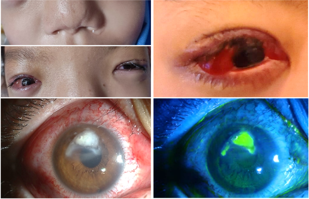
Figure 1. Clinical presentation. (A) Characteristic facial features of Noonan syndrome, including hypertelorism, a low nasal bridge, epicanthal folds, and a broad, prominent forehead. (B) Temporal bulbar conjunctival exposure beyond the palpebral fissure in the right eye. (C, D) Slit-lamp photographs showing marked conjunctival injection, chemosis, and discharge, with a 3 × 5 mm superior corneal ulcer and dense stromal infiltration extending into the pupillary zone.
Given the rapidly progressive, sight-threatening presentation, corneal scrapings were obtained immediately for microscopy and culture. Direct smear showed abundant neutrophils, occasional edematous epithelial cells, and a small number of Gram-negative bacilli. In vivo confocal microscopy demonstrated dense neutrophilic infiltration and dendritiform cells consistent with activated Langerhans cells, with no fungal hyphae or Acanthamoeba cysts identified. Bacterial culture grew P. aeruginosa after 2 days, whereas fungal culture remained negative after 5 days.
Empiric intensive antibacterial therapy was started immediately. Fortified ceftazidime (50 mg/mL) and levofloxacin 0.5% eye drops were administered alternately to the right eye every 15 minutes during the first 24 hours. Atropine ophthalmic gel was given twice daily for cycloplegia, and recombinant bovine basic fibroblast growth factor ophthalmic gel was applied to both eyes to promote ocular-surface healing. Because the ulcer had continued to enlarge despite five days of topical tobramycin and showed extensive necrosis at presentation, intravenous ceftazidime (2 g every 8 hours) was added.
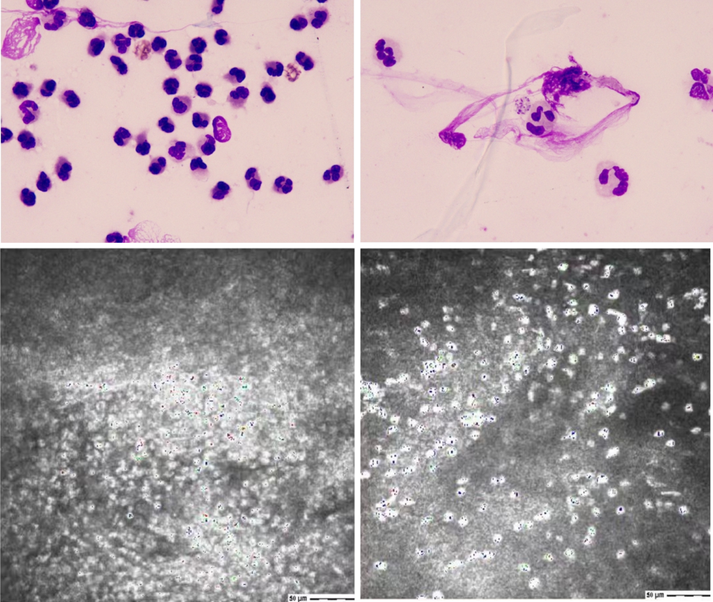
Figure 2. Microbiological and confocal examination. (A) Direct smear showing abundant neutrophils, and (B) rare Gram-negative bacilli. (C, D) In vivo confocal microscopy showing dense neutrophilic infiltration and dendritiform cells consistent with activated Langerhans cells.
By treatment day 5, conjunctival inflammation, the epithelial defect, and stromal infiltration had markedly improved. However, severe stromal thinning had developed at the site of the superior ulcer, raising concern for corneal melt (Figure 3A). As the infection continued to regress, topical antibiotics were gradually tapered, and low-dose topical corticosteroid therapy (0.02% Fluorometholone, FML) was cautiously introduced to control residual stromal inflammation. By day 10, active inflammation had largely subsided, but marked stromal thinning persisted and the risk of perforation remained (Figure 3B). Because of her congenital heart disease, any further surgery required additional perioperative assessment. Persistent eyelid edema, lagophthalmos, and ocular-surface exposure also raised concern that a corneal graft would remain vulnerable to ongoing surface stress. A bandage contact lens was therefore placed to protect the cornea. By day 21, the epithelium had largely healed, stromal inflammation had been resolved, and the cornea remained intact without perforation (Figure 3C). Visual acuity improved from counting fingers to 0.8, allowing therapeutic keratoplasty to be avoided.
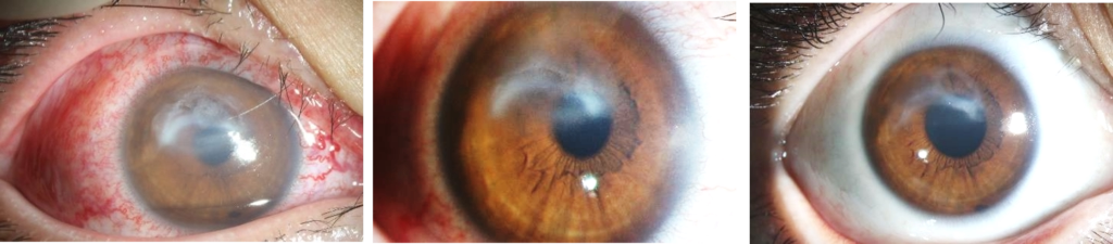
Figure 3. Treatment follow-up. (A) Day 5: marked improvement in conjunctival inflammation, epithelial defect, and stromal infiltration. (B) Day 10: inflammation largely subsided, with persistent severe stromal thinning and risk of perforation. (C) Day 21: epithelial healing with resolution of stromal inflammation and preservation of corneal integrity.
Discussion
Ptosis is a common ocular feature of Noonan syndrome, and surgical correction may further compromise eyelid closure in patients with limited baseline lid function. We report a child with Noonan syndrome who developed fulminant Pseudomonas aeruginosa keratitis shortly after bilateral ptosis surgery, progressing rapidly to severe stromal thinning and imminent perforation. This case highlights how postoperative lagophthalmos and exposure-related epithelial breakdown can create a vulnerable ocular surface and precipitate rapidly destructive corneal infection.
The rapid progression in this patient likely reflected the combined effects of postoperative exposure and Pseudomonas virulence. Noonan syndrome is associated with characteristic periocular features, including hypertelorism, epicanthal folds, altered palpebral fissure configuration, and ptosis, while ptosis surgery can further compromise eyelid closure and increase ocular surface exposure (1–6). In our patient, the superior ulcer location directly beneath the operated upper lid supported exposure-related epithelial breakdown as the initiating event rather than an intrinsic susceptibility of Noonan syndrome to Pseudomonas infection. Once the epithelial barrier was disrupted, P. aeruginosa could rapidly drive stromal destruction through bacterial proteases and an intense host inflammatory response, leading to severe thinning and an imminent risk of perforation within days (7,8). Early microbiological diagnosis and intensive antipseudomonal therapy were therefore critical to preserving corneal integrity.
Systemic ceftazidime was used as an individualized measure for rapidly progressive, necrotizing keratitis in a child for whom surgical rescue would have been high risk, rather than as routine treatment for P. aeruginosa keratitis (9). Corticosteroid therapy was likewise guided by timing: it was withheld during uncontrolled infection and introduced only after clear microbiological and clinical response, when persistent stromal damage was more likely driven by host inflammation than ongoing bacterial replication. This approach is consistent with evidence from SCUT and current guidance supporting cautious corticosteroid use after effective antimicrobial therapy has been established (10-12).
Despite marked stromal thinning, the cornea was ultimately preserved without keratoplasty. Intensive topical and systemic antimicrobial therapy rapidly controlled the infection, followed by cautious anti-inflammatory treatment after microbiological control had been achieved. By day 21, the epithelium had healed and the cornea remained intact, with substantial recovery of visual acuity. This favorable outcome emphasizes the importance of early recognition, aggressive antimicrobial treatment, and close monitoring of corneal integrity in patients who develop postoperative exposure keratopathy after ptosis surgery (13-14).
In conclusion, ptosis surgery in syndromic patients should be planned with preservation of eyelid closure in mind, rather than eyelid height alone, with careful preoperative assessment of blink, Bell phenomenon, tear film, and corneal staining. When severe keratitis develops, persistent stromal thinning after microbial control should prompt consideration of ongoing inflammation rather than immediate surgical intervention, particularly when the infiltrate is regressing, the globe remains intact, and the underlying exposure has been corrected.
Patient Consent
Written informed consent for publication of the clinical details and ophthalmic images was obtained from the patient’s parent or legal guardian.
Ethics Statement
The study was conducted in accordance with the tenets of the Declaration of Helsinki.
Conflict of Interest
The authors declare no conflicts of interest.
Funding
None
References
- van Trier DC, Vos AMC, Draaijer RW, van der Burgt I, Draaisma JMT, Cruysberg JRM. Ocular manifestations of Noonan syndrome: a prospective clinical and genetic study of 25 patients. Ophthalmology. 2016;123(10):2137–2146. doi:10.1016/j.ophtha.2016.06.061.
- Lee NB, Kelly L, Sharland M. Ocular manifestations of Noonan syndrome. Eye (Lond). 1992;6(Pt 3):328–334. doi:10.1038/eye.1992.66.
- van Trier DC, et al. Ocular findings in Noonan syndrome: a retrospective cohort study of 105 patients. Eur J Pediatr. 2018.
- van Sorge AJ, Devogelaere T, Sotodeh M, Wubbels R, Paridaens D. Exposure keratopathy following silicone frontalis suspension in adult neuro- and myogenic ptosis. Acta Ophthalmol. 2012;90(2):188–192. doi:10.1111/j.1755-3768.2010.01876.x.
- Maalouf T, George JL, et al. Serious corneal complication after ptosis surgery: six case reports. J Fr Ophtalmol. 2007;30(9):893–898.
- Young SM, et al. Lagophthalmos after congenital ptosis surgery: comparison between maximal levator resection and frontalis sling operation. Eye (Lond). 2021.
- Shin J, Son WY, Cho KJ, Rho CR. Bilateral Trichosporon asahii keratitis after ptosis correction: a case report. Medicine (Baltimore). 2021;100(29). doi:10.1097/ MD. 0000000000026688.
- O’Callaghan R, Caballero A, Tang A, Bierdeman M. Pseudomonas aeruginosa keratitis: Protease IV and PASP as corneal virulence mediators. Microorganisms. 2019;7(9):281. doi:10.3390/microorganisms7090281.
- Rhee MK, Ahmad S, Amescua G, et al.; American Academy of Ophthalmology Preferred Practice Pattern Cornea/External Disease Panel. Bacterial Keratitis Preferred Practice Pattern®. Ophthalmology. 2024;131(4)–P133. doi:10.1016/j.ophtha.2023.12.035.
- Robinson A, Kremer I, Avisar R, Gaton D, Savir H, Yassur Y. The combination of topical ceftazidime and aminoglycosides in the treatment of refractory pseudomonal keratitis. Graefes Arch Clin Exp Ophthalmol. 1999;237(3):177–180. doi:10.1007/s004170050215.
- Srinivasan M, Mascarenhas J, Rajaraman R, et al.; Steroids for Corneal Ulcers Trial Group. Corticosteroids for bacterial keratitis: the Steroids for Corneal Ulcers Trial (SCUT). Arch Ophthalmol. 2012;130(2):143–150. doi:10.1001/archophthalmol.2011.315.
- Sy A, Srinivasan M, Mascarenhas J, et al. Pseudomonas aeruginosa keratitis: outcomes and response to corticosteroid treatment. Invest Ophthalmol Vis Sci. 2012;53(1):267–272. doi:10.1167/iovs.11-7840.
- Limaiem R, Chebil A, Baba A, Ben Youssef N, Mghaieth F, El Matri L. Pediatric penetrating keratoplasty: indications and outcomes. Transplant Proc. 2011;43(2):649–651. doi:10.1016/j.transproceed.2011.01.055.
- Tzamalis A, Romano V, Cheeseman R, et al. Bandage contact lens and topical steroids are risk factors for the development of microbial keratitis after epithelium-off corneal cross-linking. BMJ Open Ophthalmol. 2019;4(1). doi:10.1136/bmjophth-2018-000231.