
Junqi Liang1-2, Zijun Zhang2, Ahyan Ilman Qudsi2
1 University College London Hospital, London, United Kingdom
2 Beijing Institute of Ophthalmology, Beijing Tongren Eye Center, Beijing Tongren Hospital, Capital Medical University, Beijing Key Laboratory of Ophthalmology and Visual Sciences, Beijing, 100005, China
Abstract
Klebsiella pneumoniae keratitis is uncommon but may progress rapidly in immunocompromised patients, leading to severe corneal destruction. We report a 42-year-old man with a history of lymphoma, hematopoietic stem cell transplantation, and chronic graft-versus-host disease who developed progressive keratitis after previous corneal ulcer repair. On presentation, visual acuity in the affected eye was hand motions, with a deep corneal ulcer, proliferative lesion, and peripheral neovascularization. Corneal culture grew K. pneumoniae. Despite intensive antimicrobial therapy, the patient required penetrating keratoplasty, anterior chamber reconstruction, and subsequent corneal repair procedures. The infection was ultimately controlled. This case highlights the increased risk of aggressive infectious keratitis in patients with post-transplant immune dysfunction and chronic ocular surface disease, and underscores the importance of early microbiological diagnosis, targeted antimicrobial therapy, and timely surgical intervention.
Introduction
Infectious keratitis is a sight-threatening corneal infection that can rapidly progress to stromal melting, perforation, and irreversible visual loss. Klebsiella pneumoniae is an uncommon but potentially destructive cause of bacterial keratitis, diagnosed through clinical assessment supported by microbiological testing of corneal specimens. Previous reports have identified prior corneal surgery or trauma, chronic ocular surface disease, and systemic immunocompromise as important predisposing factors, with severe cases often requiring therapeutic keratoplasty or other surgical intervention (1-2). We report a case of severe K. pneumoniae keratitis in a patient with chronic graft-versus-host disease following hematopoietic stem cell transplantation, highlighting the diagnostic and therapeutic challenges of aggressive corneal infection in an immunocompromised host. This report was prepared in accordance with the CARE guidelines.
Case Report
A 42-year-old man with a 5-year history of lymphoma underwent allogeneic hematopoietic stem cell transplantation 1 year before presentation and subsequently developed chronic graft-versus-host disease (cGVHD). His medical history also included gastric surgery and oral malignancy. Previous ophthalmic records documented bilateral meibomian gland orifice obstruction, trichiasis, and consistent with chronic ocular surface dysfunction. Three months before admission, he developed eye redness, pain, tearing, progressive visual loss and corneal ulcer in the right eye without an apparent trigger. He was diagnosed with infectious keratitis at a local hospital, although the exact etiology was not documented, and underwent corneal lesion debridement with amniotic membrane transplantation. Despite treatment, his ocular pain and visual symptoms continued to worsen, prompting admission to our hospital.
On presentation, visual acuity was hand motions in the right eye and 0.25 in the left eye, with intraocular pressures of 12 and 15 mmHg, respectively. Slit-lamp examination of both eyes showed rounded lid margins, trichiasis, mixed conjunctival injection, and copious ocular discharge. In the right eye, a large superior corneal infiltrate with deep ulceration and peripheral corneal neovascularization was present (Figure 1). Only the peripheral iris was faintly visible, and the remaining intraocular structures could not be visualized.
Corneal specimens were obtained for bacterial and fungal cultures, Acanthamoeba testing, and pathogen-specific PCR. Bacterial culture grew Klebsiella pneumoniae, whereas fungal and Acanthamoeba investigations were negative. Laboratory tests showed a white blood cell count of 10.9 × 10⁹/L, an absolute neutrophil count of 7.5 × 10⁹/L, and a C-reactive protein level of 33.2 mg/L.
After admission, intensive topical antimicrobial therapy was initiated with levofloxacin and tobramycin eye drops administered hourly for 2 days. Despite treatment, the corneal ulcer progressed, with increasing stromal involvement and marked hypopyon. Given the rapid clinical deterioration and extensive corneal destruction, therapeutic penetrating keratoplasty with anterior chamber irrigation and reconstruction was performed under general anesthesia. A 9.75-mm donor graft was secured with 16 interrupted 10-0 nylon sutures, and the anterior chamber was irrigated with vancomycin. By postoperative day 3, the purulent discharge had markedly decreased, and graft edema had improved, although visual acuity remained hand motions and intraocular pressure was 12 mmHg. Subsequent graft–host wound instability and eyelid malposition required further surgical intervention. One month later, the patient underwent corneal wound repair and suture correction of noncicatricial entropion. Loose inferior graft sutures were removed and replaced with three reinforcing interrupted sutures, followed by placement of a 0.5 × 0.7 cm free conjunctival flap over the corneal surface. Meanwhile, bilateral ocular GVHD was managed with topical cyclosporine, corticosteroid eye drops, and artificial tears to maintain ocular surface stability. At the 3-month follow-up, visual acuity was 0.05 in the right eye and 0.5 in the left eye, with intraocular pressures of 16 and 18 mmHg, respectively. Both ocular surfaces remained stable, with only mild corneal graft edema in the right eye (Figure 1).
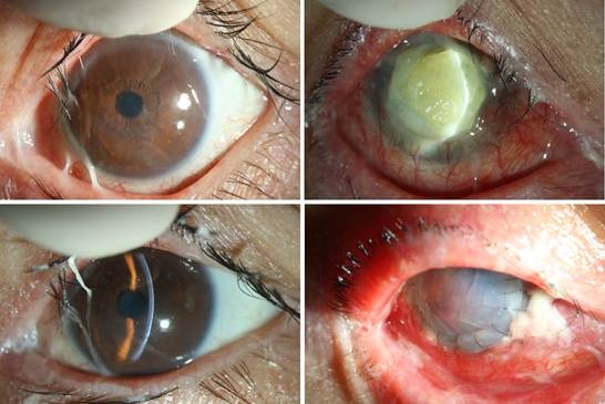
Figure 1. Slit-lamp findings in both eyes of a patient with chronic ocular graft-versus-host disease. (Top Left) Left eye showing meibomian gland orifice obstruction, mixed conjunctival injection, and copious ocular discharge. (Top Right) Right eye showing extensive yellow-white corneal infiltration and ulceration with marked conjunctival hyperemia. Following treatment of bilateral ocular GVHD with topical therapy, both ocular surfaces became stable (Bottom Row), with only mild corneal graft edema remaining in the right eye (Bottom Right).
Discussion
Ocular graft-versus-host disease (oGVHD) can disrupt multiple components of the ocular surface, including the lacrimal functional unit, meibomian glands, conjunctiva, and corneal epithelium, resulting in tear-film instability and persistent epithelial barrier dysfunction (3,4). In this patient, meibomian gland obstruction, lid-margin abnormalities, trichiasis, and bilateral epithelial disease provided additional evidence of a chronically compromised ocular surface. In a recent cohort of 173 patients with chronic oGVHD, previous ocular surgery and eyelid-margin abnormalities were independently associated with corneal ulceration, with hazard ratios of 9.16 and 3.44, respectively. Among patients who developed corneal ulcers, approximately two-thirds were infectious, and infectious ulcers were substantially more likely to result in treatment failure—defined as perforation or the need for keratoplasty—than noninfectious ulcers (57.1% vs 20.0%) (5). These features were also present in our patient, who developed fulminant infectious keratitis requiring therapeutic keratoplasty.
Klebsiella pneumoniae is an uncommon cause of microbial keratitis but can be highly destructive in susceptible eyes. The largest published series specifically examining Klebsiella keratitis identified 38 culture-proven episodes; previous keratoplasty, ocular trauma, ocular surface disease, and systemic disease were prominent predisposing factors. Corneal thinning occurred in 20 eyes, perforation in four, and 21 of 38 episodes ultimately required one or more surgical or tectonic procedures (1). Thus, although K. pneumoniae is rarely encountered as a primary corneal pathogen, its isolation in a structurally and immunologically compromised eye should not be regarded as incidental.
Microbiological confirmation is essential in severe or atypical keratitis, particularly with stromal melting, postoperative onset, or poor response to empirical therapy, because clinical appearance alone cannot reliably distinguish bacterial infection from fungal, amoebic, or sterile inflammatory disease (6). The broad work-up in our patient, including bacterial and fungal cultures, Acanthamoeba testing, and pathogen-specific PCR – was therefore appropriate. Despite intensive topical antimicrobial therapy, the ulcer continued to enlarge and developed marked hypopyon, indicating failure of medical treatment to arrest tissue destruction. Therapeutic penetrating keratoplasty was therefore undertaken primarily as an infection-eradicating and globe-preserving procedure rather than an optical intervention. Its aims first to remove infected and necrotic tissue and restore structural integrity, while visual rehabilitation is secondary. Contemporary evidence indicates that therapeutic keratoplasty can achieve infection control and globe preservation in most advanced microbial keratitis, although graft survival and final vision remain strongly influenced by disease severity, graft size, inflammation, and the condition of the recipient ocular surface (7).
The subsequent course further demonstrates that microbiological control does not equate to ocular surface recovery. After the initial infection was controlled, the patient required additional treatment for graft-host wound instability, eyelid malposition, loose sutures removing, and persistent surface compromise. Persistent epithelial defects themselves increase the risks of recurrent infection, stromal ulceration, perforation, and graft failure (8). In oGVHD, this vulnerability is amplified by tear-film deficiency, lid-margin disease, chronic inflammation, and impaired epithelial healing. Postoperative management should address the ocular surface as a functional unit rather than focus solely on pathogen eradication. In this patient, conjunctival flap coverage provided vascularized support, mechanical protection, and ocular surface stability in the setting of persistent epithelial defects, stromal thinning, and compromised tectonic integrity. (9). This case has several limitations. Detailed records from the referring hospital and the complete antimicrobial susceptibility profile of the K. pneumoniae isolate were unavailable. In addition, follow-up was relatively short, limiting assessment of long-term graft survival, recurrent infection, and rejection.
In conclusion, K. pneumoniae keratitis may progress rapidly in eyes compromised by systemic immunosuppression, chronic ocular surface disease, and prior corneal surgery. In patients with oGVHD, lid-margin disease and epithelial breakdown should be recognized as important risk factors for severe infection. Early microbiological diagnosis, targeted antimicrobial therapy, and timely therapeutic keratoplasty are essential when infection progresses or corneal integrity is threatened. Long-term success also depends on restoring and maintaining ocular surface stability to preserve both the eyeball and the graft.
Patient Consent
Written informed consent for publication of the clinical details and ophthalmic images was obtained from the patient’s parent or legal guardian.
Ethics Statement
The study was conducted in accordance with the tenets of the Declaration of Helsinki.
Conflict of Interest
The authors declare no conflicts of interest.
Funding
None
References
1. Masoumi A, Zeidabadinejad H, Jafari B, Esfandiari A, Soleimani M. Infectious keratitis caused by Klebsiella spp.: predisposing factors, presentation, and management. Int Ophthalmol. 2023;43(4):1169-1173. doi:10.1007/s10792-022-02515-y
2. Chaudhary V, Sen S, Agrawal A, Jain A, Imchen MT. Ocular surface alterations in critically ill patients: An observational study. Indian J Med Res. 2026;163(1):116-121. doi:10.25259/IJMR_1710_2025.
3. Gill K, Burgos-Blasco B, Verma A, Iovieno A, Yeung SN. Long-term clinical outcomes in chronic ocular graft-versus-host disease: a retrospective cohort study. Eye (Lond). 2026;40(10):1477-1482. doi:10.1038/s41433-026-04489-y
4. Khattak EM, Seto NA, Wong CW, et al. Current Diagnosis and Management of Ocular Graft-Versus-Host Disease at a Tertiary Cancer Center. J Clin Med. 2026;15(5):1926. Published 2026 Mar 3. doi:10.3390/jcm15051926
5. Sepulveda-Beltran PA, Carletti P, Banda V, et al. Infectious and noninfectious corneal ulcers in ocular graft-versus-host disease: epidemiology, clinical characteristics, and outcomes. Am J Ophthalmol. 2024; 257:236-246. doi:10.1016/j.ajo.2023.09.018.
6. Rhee MK, Ahmad S, Amescua G, et al.; American Academy of Ophthalmology Preferred Practice Pattern Cornea/External Disease Panel. Bacterial Keratitis Preferred Practice Pattern®. Ophthalmology. 2024;131(4). doi:10.1016/j.ophtha.2023.12.035.
7. Onofrei AG, Gheorghe AG, Dascalu AM, et al. Current challenges and long-term outcomes in corneal transplantation in infectious keratitis—a systematic review. J Clin Med. 2026;15(2):871. doi:10.3390/ jcm15020871.
8. Vaidyanathan U, Hopping GC, Liu HY, et al. Persistent corneal epithelial defects: a review article. Med Hypothesis Discov Innov Ophthalmol. 2019;8(3):163-176. PMID:31598519.
9. Zemba M, Stamate AC, Tataru CP, Branisteanu DC, Balta F. Conjunctival flap surgery in the management of ocular surface disease. Exp Ther Med. 2020;20(4):3412-3416. doi:10.3892/etm.2020.8964.