
Junqi Liang1, Ahyan Ilman Qudsi2, Xinxin Lu2
1 University College London Hospital, London, United Kingdom
2 Beijing Tongren Hospital, Capital Medical University, Beijing, 100730, China
Abstract
Purpose: To describe the clinical presentation, management, and prolonged, fluctuating course of unilateral keratitis in a patient with a documented history of mpox, and to highlight the challenges of recognizing and managing delayed ocular involvement associated with mpox.
Methods: A 22-year-old man with a recorded history of mpox presented with right-eye pain and reduced vision of approximately one month’s duration. Clinical notes, anterior segment photographs, and two in vivo confocal microscopy reports were reviewed.
Results: The right eye showed conjunctival hyperemia, corneal stromal haze and edema, peripheral corneal infiltrates, and keratic precipitates. A prior diagnosis of mpox was documented in the patient’s medical records, based on characteristic oral mucocutaneous lesions and laboratory confirmation of monkeypox virus (MPXV) infection by PCR testing of a lesion specimen. In vivo confocal microscopy during the initial episode showed dendritiform inflammatory cells in the epithelium and activated keratocytes in the anterior stroma, without fungal hyphae or Acanthamoeba cysts. Following approximately two months of topical antiviral, antibacterial, and anti-inflammatory treatment, the corneal inflammation and infiltrates markedly improved, with recovery of best-corrected visual acuity (BCVA) to 0.8. Approximately eight months later, the patient developed recurrent corneal opacity and peripheral stromal infiltrates. Topical antiviral and anti-inflammatory therapy were resumed in combination with oral acyclovir, resulting in substantial regression of the stromal infiltrates and restoration of BCVA to 0.8.
Conclusions: Mpox should be considered in the differential diagnosis of prolonged or recurrent unilateral keratitis in patients with a documented history of infection, particularly when the clinical course is atypical or fluctuating. This case highlights the potential for delayed ocular involvement and the importance of careful clinical assessment for keratitis with a history of mpox.
Introduction
Mpox-associated ocular disease includes conjunctivitis, keratitis, corneal ulceration, and scarring; of 35 cases identified by a systematic literature search, at least 40% were misdiagnosed at first presentation (1). Ocular involvement appears to be uncommon; most published evidence derives from case reports, small case series, and systematic reviews (2-6). Persistent and relapsing ocular infection has been reported, including cases supported by serial ocular polymerase chain reaction (PCR) testing (7-9). In the current global outbreak, infection has occurred predominantly among men who have sex with men, including in China (10,11). We describe a young man with a recorded history of mpox and recurrent right-eye keratitis followed for approximately 8 months.
Case Report
A 22-year-old man presented to our corneal service with a one-month history of pain and decreased vision in the right eye. The ocular symptoms had developed concurrently with oral mucocutaneous lesions. A diagnosis of mpox had previously been documented in his medical records on the basis of characteristic mucocutaneous lesions involving the oral cavity and laboratory confirmation of monkeypox virus (MPXV) infection by polymerase chain reaction (PCR) testing of a lesion specimen. He was a man who has sex with men (MSM) and tested negative for human immunodeficiency virus (HIV). He denied any history of ocular surgery or trauma, rheumatoid arthritis, or other systemic connective tissue disease.
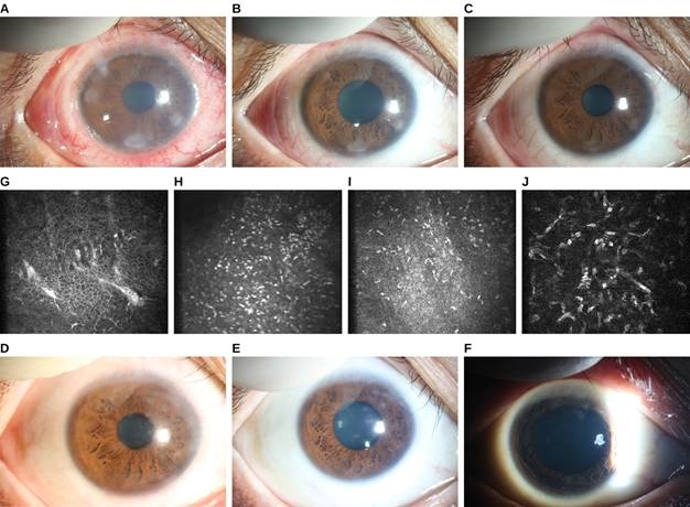
FIGURE 1. Serial right-eye photographs from six examination dates. At presentation, slit-lamp examination revealed conjunctival hyperemia and edema, wedge-shaped peripheral corneal infiltrates, central stromal edema, and keratic precipitates on the corneal endothelium (Figure 1A and 1B). In vivo confocal microscopy during the initial episode showed hyperreflective dendritic cells within the epithelium (Figure 1G), numerous hyperreflective inflammatory cells at the subepithelial level (Figure 1H and 1I), and activated keratocytes in the anterior stroma (Figure 1J). No fungal hyphae or Acanthamoeba cyst-like structures were identified. A later confocal examination again showed epithelial and stromal abnormalities.Topical treatment with ganciclovir, gatifloxacin, bovine basic fibroblast growth factor, fluorometholone 0.1%, and compound tropicamide was initiated. Conjunctival hyperemia resolved, most of the peripheral corneal infiltrates regressed. BCVA improved to 0.8 (Figure 1C).Eight months later, the patient experienced recurrent redness and blurred vision in the right eye. Examination showed patchy superficial stromal infiltrates in the peripheral cornea (Figure 1D and 1E). BCVA had decreased to 0.4. Topical antiviral and anti-inflammatory therapy were resumed, together with oral acyclovir. The ocular inflammation again improved, BCVA to 0.8 (Figure 1F).
Discussion
This case describes a prolonged, relapsing unilateral keratitis temporally associated with documented mpox. The initial episode was characterized by peripheral stromal infiltrates, stromal edema, and keratic precipitates, followed by substantial improvement and later recurrence with stromal opacity and corneal neovascularization, a recognized determinant of visual outcome in corneal disease (12,13). Similar delayed corneal inflammation has long been recognized after orthopoxvirus exposure. Disciform and interstitial keratitis, as well as iritis, have been reported after smallpox vaccination, and later vaccination-era reports have described corneal infiltrates and intraocular inflammation after vaccinia exposure (14-16). These observations support the biological plausibility of delayed or recurrent corneal inflammation after orthopoxvirus infection, but do not establish ongoing viral replication.
In this case, MPXV was confirmed from a mucocutaneous lesion, but no corneal, conjunctival, or tear specimen obtained during the ocular episodes was available for MPXV testing. The clinical phenotype is therefore not specific for MPXV. Herpetic keratitis or endotheliitis (17), postinfectious immune-mediated keratitis (18), and other inflammatory ocular surface disorders remain relevant differential diagnoses. Keratic precipitates, unilateral recurrence, and steroid responsiveness cannot by themselves distinguish among these possibilities. Published ocular mpox cases demonstrate that MPXV DNA may be detected in conjunctival swabs, corneal scrapings, or tear specimens during active ocular disease and, in some patients, again during recurrence (7-9,19,20). These observations suggest that, when ocular mpox is suspected, specimens should ideally be obtained early during an active episode and before treatment is modified (21). Nevertheless, the diagnostic performance of individual ocular specimen types has not been established, and a negative result would therefore require cautious interpretation. In vivo confocal microscopy contributed in two ways. It showed that inflammation extended from the epithelium into the anterior stroma during the initial episode (Figure 1G–J), consistent with stromal keratitis rather than ocular surface disease alone, and it identified no fungal hyphae or Acanthamoeba cyst-like structures, which argued against fungal and Acanthamoeba keratitis, both important differentials in a steroid-treated eye with stromal infiltrates.
Treatment response is similarly difficult to interpret. Improvement occurred during combined antiviral, antibacterial, and anti-inflammatory therapy, preventing attribution of benefit to any single agent. Corticosteroids may suppress immune-mediated stromal inflammation, but experimental orthopoxvirus models have also raised concern that they may impair viral clearance when active infection persists (22). Human evidence remains limited and inconsistent. Oral acyclovir administered during the later recurrence is also not evidence for an MPXV mechanism, because acyclovir has little or no established activity against orthopoxviruses (23), although its efficacy in preventing recurrent herpes simplex virus eye disease is well established (24). Conversely, clinical improvement after acyclovir does not confirm herpetic disease in the absence of virologic testing. Although not used in this case, systemic agents with anti-orthopoxvirus activity, including tecovirimat and cidofovir, have been used in severe ocular mpox (25).
The major limitations of this report are the absence of ocular MPXV testing, incomplete documentation of treatment exposure and tapering, long intervals between some examinations, and lack of standardized serial imaging, including quantitative endothelial assessment, for which measurements may differ across instruments and analytical methods.26 These limitations preclude attributing the entire course to persistent MPXV infection. Rather, the case illustrates a prolonged remitting–relapsing keratitis occurring after confirmed mpox and highlights the difficulty of distinguishing persistent viral infection from postinfectious or immune-mediated corneal inflammation on clinical morphology alone. Early ocular sampling during recurrence and longitudinal documentation of corneal findings may help clarify the mechanism in future cases.
Patient Consent
Written informed consent for publication of the clinical details and ophthalmic images was obtained from the patient’s parent or legal guardian.
Ethics Statement
The study was conducted in accordance with the tenets of the Declaration of Helsinki.
Conflict of Interest
The authors declare no conflicts of interest.
Funding
None
References
- Croasdale CR, Weinlander E, Boyce TG. Mpox keratitis: a case report and review. Cornea 2024;43:1319-1331.
- Cash-Goldwasser S, Labuda SM, McCormick DW, et al. Ocular monkeypox: United States, July-September 2022. MMWR Morb Mortal Wkly Rep 2022;71:1343-1347. doi: 10.15585/mmwr. mm7142e1
- Pazos M, Riera J, Moll-Udina A, et al. Characteristics and management of ocular involvement in individuals with monkeypox disease. Ophthalmology 2023;130:655-658.
- Curi ALL, Rojas-Carabali W, Talero-Gutiérrez C, et al. Ophthalmic manifestations of monkeypox virus infection: a case series. Ocul Immunol Inflamm 2024;32:262-265.]
- Rohilla S, Gaidhane S, Balaraman AK, et al. Ophthalmic manifestations of the mpox virus: a systematic review and meta-analysis. J Infect Dis 2025;232:220-229.
- Gandhi AP, Gupta PC, Padhi BK, et al. Ophthalmic manifestations of the monkeypox virus: a systematic review and meta-analysis. Pathogens 2023;12:452. doi: 10.3390/pathogens12030452
- Raccagni AR, Clemente T, Ranzenigo M, et al. Persistent ocular mpox infection in an immunocompetent individual. Lancet Infect Dis 2023;23:652-653.
- Pi C, Adah O, Cholli PA, et al. Relapsed mpox keratitis, St. Louis, Missouri, USA. Emerg Infect Dis 2024;30:1406-1409.
- Finamor LPS, Mendes-Correa MC, Rinkevicius M, et al. Ocular manifestations of monkeypox virus (MPXV) infection with viral persistence in ocular samples: a case series. Int J Infect Dis 2024;146:107071. doi:10.1016/j.ijid.2024.107071
- Ren R, Li C, Bai W, et al. The epidemiological characteristics of mpox cases: China, 2023. China CDC Wkly 2024;6:619-623.
- Jin Y, Qin Q, Li C, et al. Comparative analysis of epidemiological and clinical characteristics between mpox cases with and without HIV: China, 2023. China CDC Wkly 2025;7:233-238.
- Lasagni Vitar RM, Triolo G, Fonteyne P, et al. Epidemiology of corneal neovascularization and its impact on visual acuity and sensitivity: a 14-year retrospective study. Front Med (Lausanne) 2021;8:733538. doi:10.3389/fmed.2021.733538
- Bachmann B, Taylor RS, Cursiefen C. The association between corneal neovascularization and visual acuity: a systematic review. Acta Ophthalmol 2013;91:12-19.
- Semba RD. The ocular complications of smallpox and smallpox immunization. Arch Ophthalmol 2003;121:715-719. doi:10.1001/archopht.121.5.715
- Pepose JS, Margolis TP, LaRussa P, Pavan-Langston D. Ocular complications of smallpox vaccination. Am J Ophthalmol 2003;136:343-352. Doi 10.1016/s0002-9394(03)00293-9
- Fillmore GL, Ward TP, Bower KS, et al. Ocular complications in the Department of Defense Smallpox Vaccination Program. Ophthalmology 2004;111:2086-2093.
- Kaye SB, Baker K, Bonshek R, et al. Human herpesviruses in the cornea. Br J Ophthalmol 2000;84:563-571. doi:10.1136/bjo.84.5.563
- Jeng BH, Holsclaw DS. Cyclosporine A 1% eye drops for the treatment of subepithelial infiltrates after adenoviral keratoconjunctivitis. Cornea 2011;30:958-961.
- Aljawi A, Alwazae M, Babgi R, et al. Monkeypox-related keratitis: case series. Case Rep Ophthalmol 2025;16:430-438. doi:10.1159/000546007
- Mitsios A, Florou Z, Dastiridou A, et al. Monkeypox-related ophthalmic disease (MPXROD): monitoring the antiviral effect of tecovirimat with monkeypox virus detection in tear samples. Eur J Ophthalmol 2024;34:NP35-NP37.
- Centers for Disease Control and Prevention. Interim clinical considerations for management of ocular monkeypox [CDC web site]. Available at: https://www.cdc.gov/monkeypox/hcp/clinical-care/ocular-infection.html.
- Altmann S, Brandt CR, Murphy CJ, et al. Evaluation of therapeutic interventions for vaccinia virus keratitis. J Infect Dis 2011;203:683-690.
- Baker RO, Bray M, Huggins JW. Potential antiviral therapeutics for smallpox, monkeypox and other orthopoxvirus infections. Antiviral Res 2003;57:13-23.
- Herpetic Eye Disease Study Group. Acyclovir for the prevention of recurrent herpes simplex virus eye disease. N Engl J Med 1998;339:300-306. doi: 10.1056/NEJM199807303390503
- Brousse X, Kreidie R, Mourgues E, et al. Treatment of severe ocular mpox with cidofovir and tecovirimat. Emerg Infect Dis 2026;32:623-626.
- Shi Y, Huang J, Conrick P, et al. Discrepancies in endothelial cell density values of human donor corneas resulting from comparison between specular microscopes and endothelial analysis methods. Cornea 2020;39:495-500.