
Zaid Farhan1, Jonathan Than1
1Milton Keynes University Hospital NHS Foundation Trust, Milton Keynes, UK.
Summary
An 83-year-old woman with type 2 diabetes and hypertension had right central retinal vein occlusion (CRVO) and wet age-related macular degeneration, treated with intravitreal anti-VEGF injections. She was referred with a non-clearing vitreous haemorrhage of more than 6 months’ duration and a chronic superior retinal detachment; no panretinal photocoagulation was recorded before surgery.
Imaging suggested tractional retinal membranes, and 25-gauge vitrectomy found extensive fibrovascular proliferation and tractional retinal detachment. Intraoperative events included hypotony, a large posterior retinal break and a limited suprachoroidal haemorrhage. Intraocular pressure later fell (14, 6, then 3 mmHg), with a mature cataract and chronic retinal re-detachment under oil.
On the day of planned combined surgery, the surgeon judged the eye hypotonous and phthisical; after discussion, surgery was cancelled. The fellow eye had a recent CRVO (vision 6/30). The case illustrates recognising an eye that should not be operated on, and shared decision-making.
Background
Ischaemic central retinal vein occlusion (CRVO) can cause vitreous haemorrhage and tractional retinal detachment, as eyes with extensive retinal non-perfusion are at high risk of retinal neovascularisation and neovascular glaucoma (1). Intravitreal anti-vascular endothelial growth factor (anti-VEGF) therapy is NICE-recommended for macular oedema after CRVO and for wet age-related macular degeneration (AMD) (1–3). In ischaemic CRVO, however, anti-VEGF therapy may mask or delay, but not ameliorate, neovascular complications, so surveillance is recommended (1). We describe an eye in which chronic retinal detachment under silicone oil led to hypotony and impending phthisis, and the reasoning behind a decision not to operate.
Case presentation
An 83-year-old woman had type 2 diabetes, systemic hypertension on antihypertensives and arthritis, and took aspirin. She took no anticoagulants and did not smoke.
The right eye had CRVO with macular oedema and wet AMD, both treated with intravitreal anti-VEGF therapy (ranibizumab and aflibercept). Macular scarring and atrophy were noted in January 2026. Injections had been stopped, after which she developed a vitreous haemorrhage. No panretinal photocoagulation (PRP) had been performed before surgery due to poor fundal view since onset of vitreous haemorrhage. The left eye had ocular hypertension from 2022 (untreated intraocular pressure (IOP) 31 mmHg), treated with bimatoprost, then preservative-free latanoprost/timolol from January 2024.
In January 2026, a neighbouring district general hospital found a symptomatic, non-clearing vitreous haemorrhage of more than 6 months in the right eye. It also found a shallow, chronic superior retinal detachment involving the upper macula. Optical coherence tomography (OCT) demonstrated an attached fovea. After counselling about a guarded visual prognosis, she was booked for vitrectomy with gas and referred to our vitreoretinal unit (Table 1).
In February 2026, both lenses showed nuclear sclerotic opacity. The clinic diagnosis was right vitreous haemorrhage, possibly secondary to retinal vein occlusion with retinal ischaemia and retinal neovascularisation, with a guarded visual prognosis because of the macular scar. Vitrectomy with endolaser panretinal photocoagulation, with or without delamination, was planned under local anaesthesia. She was counselled about an approximately 90% chance of success and a 5 to 10% risk of rebleed. Consent listed risks including rebleeding, infection, inflammation, cataract, high or low pressure, gas or oil, retinal detachment and loss of vision.
| When | Setting | VA (right, left) | IOP (right, left; mmHg) | Event |
| Jan 2026 | Referring DGH | CF, 6/9−2 | 13, 22 | Non-clearing VH >6 months; superior chronic retinal detachment |
| Feb 2026 | Our clinic | HM, 0.22 logMAR | 14, 17 | OCT (Figure 1); vitrectomy planned |
| Mar 2026 | Theatre | — | — | 25-gauge vitrectomy, delamination, laser, silicone oil 1300 cSt |
| May 2026 | Clinic (~7 weeks) | HM, 6/7.6 | 14, 20 | Poor view; posterior subcapsular cataract; possible inferior retinal detachment under oil |
| Jun 2026 | Pre-assessment | — | — | Combined cataract surgery, removal of silicone oil, proliferative vitreoretinopathy peel, retinectomy, laser retinopexy and silicone oil injection operation booked |
| Before surgery | Second pre-operative assessment | — | 6, — | Mature (white) cataract; no fundal view; OCT suggested re-detachment under oil |
| Sep 2026 | Day of surgery | HM, 6/30 | 3, 15 | Soft globe, phthisical eye; surgery cancelled |
Table 1. Visual acuity (VA), IOP and clinical course. CF, counting fingers; HM, hand movements; DGH, district general hospital; VH, vitreous haemorrhage.
Investigations
Right-eye IOP was 13, 14, 17, 14, 6 and 3 mmHg (Table 1; Figure 3), measured by rebound tonometry in January and February 2026. We took hypotony as IOP of 5 mmHg or less (4).
OCT in February 2026 was consistent with tractional retinal detachment, with preretinal fibrovascular proliferation (Figure 1).
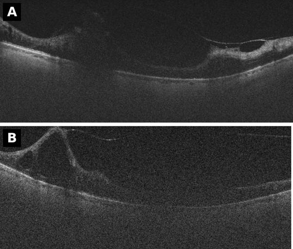
Figure 1. Right-eye macular OCT, February 2026, before vitrectomy. (A) Reduced signal from media opacity, with an intraretinal cystoid space and a preretinal fibrovascular membrane. (B) A peaked, tent-like retinal elevation with a vitreous strand inserting at its apex, consistent with vitreoretinal traction. The superior tractional retinal detachment lies outside these macular frames.
Right-eye diabetic retinopathy was “ungradable” due to vitreous haemorrhage. No neovascularisation of the iris was noted. No fluorescein angiography was performed at our unit.
In May 2026 OCT could not be acquired. At a second pre-operative assessment, IOP was 6 mmHg, the cataract was mature with no fundal view, and OCT suggested re-detachment under oil.
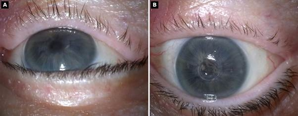
Figure 2. Right eye under the operating microscope on the day of planned surgery, September 2026 (IOP 3 mmHg). (A) Irregular corneal surface with an indentation, consistent with a soft globe and collapsed anterior chamber. (B) Small pupil with pigment and whitish lens opacity (mature cataract) in the visual axis; no red reflex or fundal view.
On the day of surgery (Figure 2), the operating surgeon found the right globe soft to digital palpation, with a corneal indentation. The eye was described as “completely hypotonous and phthisical”.
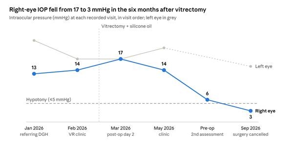
Figure 3. Right-eye intraocular pressure at each recorded visit, January to September 2026, in visit order. Dashed line: hypotony threshold of 5 mmHg. The 6 mmHg reading is from the second pre-operative assessment.
Differential diagnosis
Possible contributors to the haemorrhage and tractional retinal detachment include ischaemic CRVO (1) or proliferative diabetic retinopathy. Breakthrough bleeding from neovascular AMD could explain the vitreous haemorrhage but not the fibrovascular proliferation. In January 2026 the superior elevation was considered retinal detachment or retinoschisis.
The later hypotony was attributed to ciliary body insufficiency secondary to chronic retinal detachment under silicone oil, progressing to phthisis (5).
Treatment
In March 2026 a consultant vitreoretinal surgeon performed 25-gauge three-port vitrectomy. Adherent vitreous face and extensive fibrovascular proliferation were found; delamination was performed. A superior retinal detachment with a retinal break and subretinal haemorrhage was present. The operative note records an episode of hypotony, followed by a large superotemporal posterior retinal break with retinal detachment and a limited suprachoroidal haemorrhage. Infusion pressure was raised and no further suprachoroidal haemorrhage occurred. After subretinal fluid drainage, endolaser PRP and laser around the breaks (798 shots) were applied, followed by fluid–air exchange (retina flat) and air–silicone oil exchange (1300 cSt).
Combined cataract surgery, removal of silicone oil, proliferative vitreoretinopathy peel, retinectomy, laser retinopexy and silicone oil re-injection was booked in June 2026. On the day of surgery in September 2026, the eye was noted to have become phthisical, and in the operating surgeon’s judgement, further surgery would not improve vision and could increase inflammation and pain (high-risk). After discussion, the patient and surgeon agreed not to proceed, and the operation was cancelled. The documented plan was dexamethasone drops three times daily for inflammation or discomfort, with silicone oil left in situ. The existing oil should help prevent a cosmetically unacceptable appearance despite phthisis bulbi.
Outcome and follow-up
At the last review, the right eye had hand-movement vision, IOP 3 mmHg and silicone oil in situ; left-eye VA had fallen from 6/7.6 (May) to 6/30 (September 2026). A recent left CRVO had been treated with two anti-VEGF injections, with a third planned and PRP possible later. Low vision clinic and Eye Clinic Liaison Officer support were to be offered, with 2-week follow-up at our unit. The hypotony was attributed to ciliary body insufficiency with progression to phthisis; ongoing follow-up will continue to assess for, and if necessary address, any pain or change in cosmetic appearance.
Discussion
In this eye, chronic retinal re-detachment under silicone oil after complex surgery for tractional detachment led to ciliary body insufficiency, progressive hypotony and impending phthisis. The key decision came on the morning of surgery: a soft globe, a collapsed anterior chamber and an IOP of 3 mmHg showed that the planned operation offered little visual benefit and considerable risk, and the patient and surgeon agreed not to proceed.
The course began with ischaemic CRVO. Anti-VEGF therapy delays rather than prevents neovascular complications in ischaemic CRVO, and Royal College of Ophthalmologists guidance recommends monitoring for neovascularisation, angiography to assess non-perfusion and 1 to 2 monthly reviews in the first year after treatment stops, noting that PRP may help prevent vitreous haemorrhage (1). No PRP had been performed before surgery, and by the time of vitrectomy extensive fibrovascular proliferation had produced a tractional detachment.
Retinal detachment lowers IOP in two ways: it increases outflow through unconventional, uveoscleral pathways and it reduces aqueous production (6). Ciliary body detachment is often present but does not appear to cause the reduced aqueous formation, and control of the accompanying inflammation is needed for aqueous dynamics to recover (6), consistent with the decision to continue topical steroid. Hypotony is common after surgery for complex detachment: in the Silicone Study, chronic hypotony (5 mmHg or less on two consecutive visits or any three) affected 24% of eyes operated on for severe proliferative vitreoretinopathy (4). Our patient’s IOP fell from 14 to 6 and then 3 mmHg; only the last reading met that threshold, but 6 mmHg was already below the statistical definition of hypotony (below 6.5 mmHg) (5).
Phthisis bulbi is the end stage of this process, with shrinkage and disorganisation of the globe driven by hypotony, breakdown of the blood–ocular barrier and inflammation. Retinal detachment is a recognised cause, and once phthisis is established, management aims at comfort and appearance rather than vision (7). In our patient the signs were clinical and immediate: a globe soft to palpation, corneal indentation and a collapsed anterior chamber. Recognising them on the day, rather than proceeding on a plan made three months earlier, changed the balance of the operation.
The planned operation would have combined cataract surgery, silicone oil removal, proliferative vitreoretinopathy peel, retinectomy and oil re-injection. Proliferative vitreoretinopathy remains a significant challenge for vitreoretinal surgeons, and its incidence has not fallen despite advances in technique (8). In a UK series of 304 eyes with proliferative vitreoretinopathy managed by retinectomy, final reattachment was achieved in 72%, vision improved in 45% and worsened in 29%; reproliferation and hypotony were the main reasons for failure, and final hypotony was associated with silicone oil removal (9). Oil removal itself led to redetachment in 25%, hypotony in 16% and expulsive haemorrhage in 1% of 85 patients in an earlier UK series (10). With a macular scar and a chronically detached retina, our patient’s visual potential was minimal, so these risks would have bought little. Long-term oil retention is an accepted option in selected eyes, including those with persistent hypotony, although function is limited (40% ambulatory vision in a UK series of 50 eyes) (11). In this eye, the oil was left in situ to limit the cosmetic effect of phthisis by retaining a more physiological ocular axial length.
Further surgery also carried specific hazards. Suprachoroidal haemorrhage complicated 1.03% of 5,459 vitrectomies in a UK series; older age and antiplatelet or anticoagulant use were risk factors, and phthisis was more common afterwards (7.1% versus 0.9%) (12). A recent systematic review also lists ocular hypotony among the risk factors for perioperative suprachoroidal haemorrhage (13). Our patient was 83, took aspirin, was hypotonous and had already had a limited suprachoroidal haemorrhage during her first operation. Sympathetic ophthalmia is rare (minimum incidence 0.03 per 100,000 in UK and Irish surveillance), but 11 of 18 cases with baseline data followed surgery, six after retinal surgery alone (14), and the risk may be greater after multiple vitreoretinal procedures (15). This mattered because her left eye, now her only useful eye, has a recent CRVO under treatment; if ischaemic, more than 90% of such patients end at 6/60 or worse (1). Sympathetic ophthalmia in this fellow eye would likely have a devastating impact upon quality of life.
Consent is a continuing process, and General Medical Council guidance requires that reasonable alternatives, including taking no action, are discussed (16). The Montgomery judgment requires disclosure of material risks and reasonable alternatives (17), and NICE guidance on shared decision-making states that people should know their options include choosing no treatment (18). The day-of-surgery review was the last point at which new findings could change that discussion. Here, it led to a shared decision not to operate, with topical steroid, retention of the oil and low vision support.
Learning points
- In ischaemic CRVO, anti-VEGF therapy may delay rather than prevent neovascular complications: document ischaemia and plan regular surveillance.
- In retinal vein occlusion, a non-clearing vitreous haemorrhage can conceal fibrovascular proliferation and traction, making surgery more complex and prognosis more guarded; counsel accordingly and plan pre-operatively with B-scan ultrasonography.
- After complex vitrectomy, re-measure IOP and examine the globe at every visit; hypotony (5 mmHg or less), particularly with chronic detachment under oil, can progress to phthisis.
- Recognising a hypotonous, pre-phthisical eye on the day of surgery allows low-yield, high-risk surgery to be cancelled; not operating is an alternative to discuss.
- When the fellow eye is threatened, weigh further surgery on the poorer eye against inflammation, pain and sympathetic ophthalmia, and arrange low vision support early.
References
- The Royal College of Ophthalmologists. Retinal Vein Occlusion (RVO) Clinical Guidelines. London: RCOphth; 2022 (review date January 2025).
- National Institute for Health and Care Excellence. Ranibizumab for treating visual impairment caused by macular oedema secondary to retinal vein occlusion. TA283. London: NICE; 2013 (updated 2024 May 20).
- National Institute for Health and Care Excellence. Age-related macular degeneration. NG82. London: NICE; 2018.
- Barr CC, Lai MY, Lean JS, Linton KL, Trese M, Abrams G, et al; Silicone Study Group. Postoperative intraocular pressure abnormalities in the Silicone Study. Silicone Study Report 4. Ophthalmology 1993;100(11):1629-35. doi:10.1016/S0161-6420(93)31425-9
- Thomas M, Vajaranant TS, Aref AA. Hypotony maculopathy: clinical presentation and therapeutic methods. Ophthalmol Ther 2015;4(2):79-88. doi:10.1007/s40123-015-0037-z
- Pederson JE. Ocular hypotony. Trans Ophthalmol Soc U K 1986;105(Pt 2):220-6.
- Tripathy K, Chawla R, Temkar S, Sagar P, Kashyap S, Pushker N, et al. Phthisis bulbi—a clinicopathological perspective. Semin Ophthalmol 2018;33(6):788-803. doi:10.1080/08820538.2018.1477966
- Charteris DG. Proliferative vitreoretinopathy: revised concepts of pathogenesis and adjunctive treatment. Eye (Lond) 2020;34(2):241-5. doi:10.1038/s41433-019-0699-1
- Grigoropoulos VG, Benson S, Bunce C, Charteris DG. Functional outcome and prognostic factors in 304 eyes managed by retinectomy. Graefes Arch Clin Exp Ophthalmol 2007;245(5):641-9. doi:10.1007/s00417-006-0479-z
- Casswell AG, Gregor ZJ. Silicone oil removal. II. Operative and postoperative complications. Br J Ophthalmol 1987;71(12):898-902. doi:10.1136/bjo.71.12.898
- Morphis G, Irigoyen C, Eleuteri A, Stappler T, Pearce I, Heimann H. Retrospective review of 50 eyes with long-term silicone oil tamponade for more than 12 months. Graefes Arch Clin Exp Ophthalmol 2012;250(5):645-52. doi:10.1007/s00417-011-1873-8
- Chandra A, Xing W, Kadhim MR, Williamson TH. Suprachoroidal hemorrhage in pars plana vitrectomy: risk factors and outcomes over 10 years. Ophthalmology 2014;121(1):311-7. doi:10.1016/j.ophtha.2013.06.021
- Ribeiro M, Monteiro DM, Moleiro AF, Rocha-Sousa A. Perioperative suprachoroidal hemorrhage and its surgical management: a systematic review. Int J Retina Vitreous 2024;10(1):55. doi:10.1186/s40942-024-00577-x
- Kilmartin DJ, Dick AD, Forrester JV. Prospective surveillance of sympathetic ophthalmia in the UK and Republic of Ireland. Br J Ophthalmol 2000;84(3):259-63. doi:10.1136/bjo.84.3.259
- Ullrich K, Patel BCK, Malhotra R. Material risk: vitreoretinal surgery, evisceration, enucleation and sympathetic ophthalmia—where are we currently? Eye (Lond) 2023;37(17):3542-50. doi:10.1038/s41433-023-02562-4
- General Medical Council. Decision making and consent. London: GMC; 2020.
- Montgomery v Lanarkshire Health Board (2015) UKSC 11.
- National Institute for Health and Care Excellence. Shared decision making. NG197. London: NICE; 2021 (updated 2021 Oct).