
Junqi Liang 1-2, Ahyan Ilman Qudsi 2, Xinxin Lu 2
1 University College London Hospital, London, United Kingdom
2 Beijing Tongren Hospital, Capital Medical University, Beijing, 100730, China
Abstract
Purpose: To report a case of multidrug-resistant Corynebacterium amycolatum and Aspergillus flavus keratitis following conjunctival flap surgery before microbiological diagnosis.
Observations: A 77-year-old man with diabetes rubbed his right eye during farm work. Six days later, he was diagnosed with fungal keratitis at a local hospital, where a conjunctival flap was sutured over the cornea. At referral, a dense infiltrate was noted beneath the flap, and in vivo confocal microscopy (IVCM) revealed filamentous structures suggestive of fungal hyphae. A corneal swab grew a moderate quantity of C. amycolatum, resistant to penicillin, ceftriaxone, erythromycin, levofloxacin, gatifloxacin and chloramphenicol and susceptible only to vancomycin and rifampicin, with a scant A. flavus. Cytology showed a neutrophil-predominant infiltrate. On empirical therapy that included levofloxacin, vision fell to light perception. After levofloxacin was replaced with rifampicin and the flap sutures were removed, the retracting flap exposed an ulcer with a 3-mm hypopyon. Serial IVCM showed dense stromal inflammation with few identifiable hyphae. After the flap retracted and tobramycin was added, topical natamycin and chlorhexidine were continued. The hypopyon resolved within 8 days, and visual acuity improved to 20/400 at 8 weeks without keratoplasty.
Conclusions and importance: C. amycolatum should not be dismissed as a commensal when it grows from a corneal ulcer with a leukocyte response, even alongside a fungus. This isolate resisted the fluoroquinolones and the third-generation cephalosporin given empirically, and only species identification and susceptibility testing pointed to active drugs.
Introduction
Corynebacterium species are common ocular surface flora and can easily be dismissed as contaminants when recovered from corneal specimens (1,2). In 1989 Rubinfeld and colleagues argued that these diphtheroids can be true ocular pathogens (3). At our institution, Gram-positive bacilli, most of them Corynebacterium, rose from 4.2% to 16.1% of bacterial keratitis isolates between the periods before and after the COVID-19 pandemic (4). Corynebacterium amycolatum is among the coryneforms most often isolated from clinical samples, but its variable biochemistry leads to misidentification, and individual reports of C. amycolatum keratitis remain scarce (5-7).
We describe a patient whose cornea was infected by multidrug-resistant C. amycolatum together with Aspergillus flavus. A conjunctival flap placed before microbiological diagnosis concealed the course of the infection, and serial in vivo confocal microscopy (IVCM) recorded the cornea through the flap and after it retracted.
Case report
A 77-year-old man, a farmer, rubbed his right eye while working in the fields and developed redness, foreign-body sensation, discharge and blurred vision. He had had diabetes mellitus for more than 10 years and a cerebral infarction 12 years earlier. Six days after onset, an outside hospital diagnosed fungal keratitis and sutured a partial conjunctival flap over the right cornea. He was referred to our eye center the next day, designated day 1. At the initial examination, a hemorrhagic flap covered most of the corneal surface in the right eye. Beneath the flap was a dense, white infiltrate with feathery margins extending to the inferior limbus (Figure 1A). Corneal scrapings were taken for cytology and corneal swabs for bacterial and fungal culture from the infiltrate exposed at the flap margin. Cytology showed a neutrophil-predominant infiltrate with degenerating neutrophils, epithelial cells and erythrocytes (Figure 1G–I). The bacterial culture grew a moderate quantity of C. amycolatum, identified by MALDI-TOF mass spectrometry. On disk diffusion testing the isolate resisted penicillin, ceftriaxone, erythromycin, levofloxacin, gatifloxacin and chloramphenicol and was susceptible to vancomycin and rifampicin (Table 1). The fungal culture grew a scant quantity of A. flavus. IVCM imaging through the flap showed a hazy field with dense small round cells and fine, interlacing, branching filaments suggestive of hyphae (Figure 2A). Therefore, oral voriconazole (100 mg twice daily) and topical natamycin (5%), chlorhexidine (0.02%), and levofloxacin (0.5%) were initiated on day 2.
On day 7, visual acuity had fallen to light perception, the eyelids and conjunctiva were swollen, and a large grayish-white infiltrate occupied the cornea beneath the flap. Based on the antimicrobial susceptibility testing results, levofloxacin was replaced with topical rifampicin 0.1%. On day 14 the flap sutures were removed under the operating microscope, and visual acuity was hand motions. Hyperreflective inflammatory cells infiltrated it to approximately 190 µm, and no hyphae were clearly identified with IVCM examination (Figure 2B). By day 21 the flap had retracted toward the limbus, exposing a central ulcer with a 3-mm hypopyon, and sheets of round inflammatory cells in the superficial ulcer bed (Figure 1B, 1C and 2C). Topical tobramycin 0.3% and rifampicin 0.1%, each five times daily, and a single 2-g intravenous dose of ceftriaxone were added for one week. The eye then improved. On day 28 the ulcer was smaller and the hypopyon had resolved. The inflammatory cells were fewer, fragmented hyperreflective aggregates remained in the anterior stroma, and discrete deposits lay on the endothelium (Figures 1D, 2D and 2E). On day 42 visual acuity was 20/2000 and intraocular pressure 7.9 mmHg, with stromal edema and pigmented keratic precipitates. Only mature dendritic cells were detected, and the endothelial mosaic was preserved apart from folds (Figures 1E, 1F and 2F–H). Natamycin was reduced to four times daily, and bromfenac 0.1% and carbomer gel were added. On day 56 visual acuity was 20/400 and the central infiltrate had largely regressed, leaving an irregular epithelium, mild stromal edema, posterior synechiae and cataract. By then rifampicin and tobramycin 0.3% had been stopped, the globe was preserved without keratoplasty, and follow-up continues.
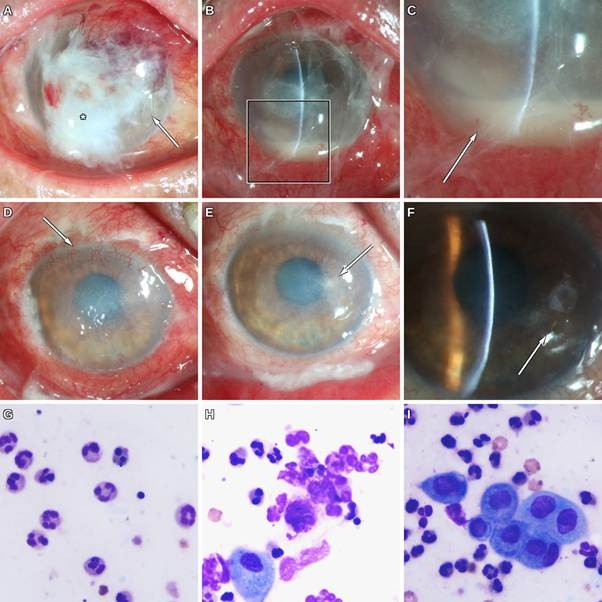
Figure 1. Clinical course and cytology of the corneal scraping in the right eye. (A) Day 1. A conjunctival flap covers most of the cornea, with a suture visible (arrow) and a dense white infiltrate beneath the flap (asterisk). (B) Day 21. The flap has retracted, exposing a central stromal infiltrate with edema; the boxed area is enlarged in Figure 1C. (C) Hypopyon in the anterior chamber (arrow). (D) Day 28. The hypopyon has resolved; stromal haze persists, and superficial vessels extend from the superior limbus (arrow). (E-F) Day 42. The cornea is clearer, with a residual paracentral stromal opacity (arrow) and pigmented keratic precipitates (arrow). (G–I) Cytology of the corneal scraping on day 1 (Giemsa stain, ×1000): (G) neutrophil-predominant infiltrate; (H) degenerating neutrophils and necrotic debris, with an epithelial cell; (I) epithelial cells surrounded by neutrophils, with scattered erythrocytes.
Table 1. Disk diffusion susceptibility of the Corynebacterium amycolatum isolate.
| Agent | Zone diameter (mm) | Interpretation | S / I / R (Laboratory criteria, mm) |
| Penicillin | 6 | R | ≥29 / – / ≤28 |
| Ceftriaxone | 6 | R | ≥23 / 20–22 / ≤19 |
| Erythromycin | 6 | R | ≥23 / 14–22 / ≤13 |
| Levofloxacin | 6 | R | ≥24 / 21–23 / ≤20 |
| Gatifloxacin | 10 | R | ≥23 / 20–22 / ≤19 |
| Chloramphenicol | 6 | R | ≥18 / 13–17 / ≤12 |
| Vancomycin | 22 | S | ≥17 / 15–16 / ≤14 |
| Rifampicin | 33 | S | ≥20 / 17–19 / ≤16 |
Note: A zone of 6 mm equals the disk diameter, meaning no inhibition. Criteria are those applied by the testing laboratory; CLSI M45 does not provide disk diffusion criteria for Corynebacterium species. I, intermediate; R, resistant; S, susceptible.
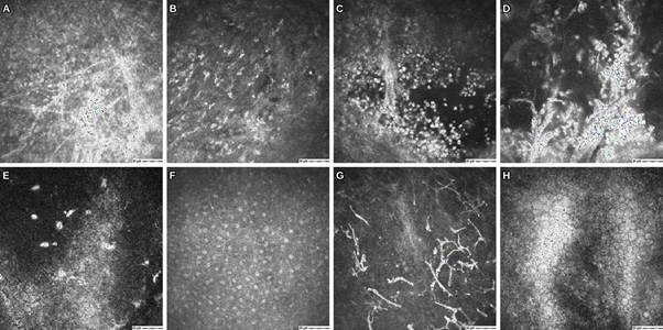
Figure 2. Serial in vivo confocal microscopy of the right cornea. (A) Day 2, through the conjunctival flap: fine interlacing branching filaments in a hazy field. (B) Day 14, anterior stroma (67 µm): dense hyperreflective inflammatory cells. (C) Day 21, superficial ulcer bed: round inflammatory cells. (D-E) Day 28, anterior stroma (64 µm): clustered hyperreflective aggregates with fewer inflammatory cells and discrete hyperreflective endothelial deposits. (F-H) Day 42, epithelium and Bowman layer: branching hyperreflective structures consistent with mature dendritic cells; endothelium: endothelial mosaic with folds. (400 × 400 µm; scale bar, 50 µm).
Discussion
The fungal diagnosis made at the outside hospital was correct but incomplete. Although A. flavus was isolated, multidrug-resistant C. amycolatum was recovered in greater quantity. The conjunctival flap placed on the day of diagnosis masked the co-infection until it retracted nearly 3 weeks later, revealing an ulcer with hypopyon. Because C. amycolatum colonizes skin and mucosa, its role has to be argued. Sugumaran et al. required repeat isolation in pure culture and a leukocyte reaction on direct microscopy before accepting it as the cause of a corneal ulcer (6). Our isolate came from a single specimen, which grew moderate amounts of C. amycolatum and A. flavus. Meanwhile, cytology of corneal scrapings showed a neutrophil-predominant infiltrate with degenerating neutrophils, providing the required leukocyte reaction. Two additional findings further support a causal role.
Species identification changed the choice of drug. In a Japanese series dominated by Corynebacterium macginleyi, 91% of corneal isolates resisted fluoroquinolones but none resisted cefmenoxime, and the authors recommended topical cephalosporins when Corynebacterium keratitis is suspected (8). That advice may not hold for C. amycolatum, which is frequently resistant to beta-lactams (9); our isolate resisted both penicillin and ceftriaxone. Fluoroquinolone resistance among corneal Corynebacterium isolates ranged from 45% to 58% across three Indian centers (7), and at our institution gatifloxacin resistance in Corynebacterium rose from 22.2% before the pandemic to 46.4% after it (4). Corynebacteria lack topoisomerase IV, so mutations in DNA gyrase alone are enough to confer resistance (8,10). Vancomycin was active against every isolate in the two most recent series (7,8) and was part of the regimen that healed the earlier C. amycolatum ulcer (6). Topical rifampicin, which is available in China, was the other active agent here; where it is not, fortified vancomycin is the reasonable alternative. Our susceptibility data carry a caveat, because Clinical and Laboratory Standards Institute document M45 gives interpretive criteria for Corynebacterium based on minimum inhibitory concentrations rather than disk diffusion (6,11). Five agents produced no zone of inhibition and gatifloxacin a 10-mm zone, so those resistance calls do not depend on the breakpoints chosen; the vancomycin and rifampicin zones were 22 and 33 mm.
Mixed infection is common in Corynebacterium keratitis. Soni et al. found it in 43 of 108 cases; a fungus was the co-pathogen in 22, and A. flavus grew in two, although the Corynebacterium species paired with each fungus was not reported (7). Compared with fungal keratitis alone, polymicrobial keratitis affects older patients, presents with larger infiltrates and is more often complicated by endophthalmitis (12), and its outcomes are worse than those of monomicrobial disease (13). Our patient, aged 77 with diabetes and an infiltrate reaching the limbus, fits that pattern. Within Corynebacterium keratitis, however, Soni et al. found no difference in outcome between pure and mixed infection (7).
Gundersen introduced his conjunctival flap technique for chronic corneal disease in 1958 (14), and it is still used when the ocular surface fails to heal (15). Chinese centers use partial or total flaps as a transitional operation for refractory fungal keratitis (16,17). These series used the flap for refractory or severe disease, and Sun et al. operated only after more than a week of topical and systemic antifungal treatment had failed, removed the necrotic tissue first and used ultrasonography to exclude endophthalmitis (18). Our patient received a flap on the day fungal keratitis was diagnosed, before any culture result. Beneath the flap, IVCM images lost contrast and depth reference, and the hypopyon was first recorded after the flap retracted. The eye improved within 8 days of the flap being found retracted, which fits, but does not prove, impaired drug penetration beneath it. Its sutures may also have offered a surface for the bacterium, since Corynebacterium macginleyi forms biofilms on corneal sutures (19). IVCM detects fungal hyphae with high sensitivity in experienced hands (20,21), and Aspergillus hyphae have a recognizable branching pattern (22). It cannot resolve bacteria. A positive IVCM result for hyphae should not replace bacterial culture. Dendritic cells increase in number and size, with longer dendrites, in bacterial and fungal keratitis (23), and we interpreted the branching structures seen on day 42 in that light.
This report has some limits. Cultures were not repeated, susceptibility was tested by disk diffusion, and treatment changed several times, so no single drug can be credited with the improvement. The oral voriconazole dose was low. The posterior segment was not assessed by B-scan ultrasonography. Follow-up so far is 8 weeks.
Conclusions
C. amycolatum can act as a corneal pathogen together with A. flavus in an older patient with diabetes after agricultural trauma. Because the isolate resisted fluoroquinolones and a third-generation cephalosporin, only species identification and susceptibility testing identified effective drugs. A conjunctival flap placed before microbiological diagnosis concealed progression in this eye and is better deferred until culture results are available.
Patient consent
Written informed consent to publish this case report and the accompanying images was obtained from the patient.
Funding
This research did not receive any specific grant from funding agencies in the public, commercial, or not-for-profit sectors.
Declaration of competing interest
The authors declare that they have no known competing financial interests or personal relationships that could have appeared to influence the work reported in this paper.
Authorship
All authors attest that they meet the current ICMJE criteria for authorship.
Declaration of generative AI and AI-assisted technologies in the manuscript preparation process
During the preparation of this work the authors used Claude (Anthropic) in order to assist with language editing. After using this tool, the authors reviewed and edited the content as needed and take full responsibility for the content of the published article.
Acknowledgments
We thank the Ocular Microbiology Laboratory of Beijing Tongren Hospital for culture and susceptibility testing.
References
1. Aoki T, Kitazawa K, Deguchi H, Sotozono C. Current evidence for Corynebacterium on the ocular surface. Microorganisms 2021;9:254. https://doi.org/10.3390/microorganisms9020254.
2. Das S, Rao AS, Sahu SK, Sharma S. Corynebacterium spp as causative agents of microbial keratitis. Br J Ophthalmol 2016;100:939-43. https://doi.org/10.1136/bjophthalmol-2015-306749.
3. Rubinfeld RS, Cohen EJ, Arentsen JJ, Laibson PR. Diphtheroids as ocular pathogens. Am J Ophthalmol 1989;108:251-4. https://doi.org/10.1016/0002-9394(89)90114-1
4. Shi Q, Mao D, Zhang Z, Qudsi AI, Wei M, Cheng Z, et al. Epidemiological and antimicrobial resistance trends in bacterial keratitis: a hospital-based 10-year study (2014-2024). Microorganisms 2025;13:670. https://doi.org/10.3390/microorganisms13030670.
5. Funke G, von Graevenitz A, Clarridge JE 3rd, Bernard KA. Clinical microbiology of coryneform bacteria. Clin Microbiol Rev 1997;10:125-59. https://doi.org/10.1128/CMR.10.1.125.
6. Sugumaran R, Sistla S, Chavhan P, Deb AK. Corynebacterium amycolatum: an unusual cause of corneal ulcer. BMJ Case Rep 2020;13:e237818. https://doi.org/10.1136/bcr-2020-237818.
7. Soni T, Gunasekaran R, Das S, Mitra S, Prajna NV, Prajna L, et al. Corynebacterium keratitis: pure versus mixed infection and antibiotic susceptibility patterns from different tertiary eye care centers. Cornea 2022;41:1386-91. https://doi.org/10.1097/ICO.0000000000002915.
8. Inoue H, Toriyama K, Murakami S, Miyamoto H, Ikegawa W, Takezawa Y, et al. Clinical and bacteriological characteristics of Corynebacterium keratitis. J Ophthalmic Inflamm Infect 2025;15:2. https://doi.org/10.1186
9. Zalas P, Mikucka A, Gospodarek E. [Antibiotic sensitivity of Corynebacterium amycolatum]. Medycyna Doswiadczalna i Mikrobiologia. 2004 ;56(4):327-334. PMID: 15959988.
10. Eguchi H, Kuwahara T, Miyamoto T, Nakayama-Imaohji H, Ichimura M, Hayashi T, Shiota H. High-level fluoroquinolone resistance in ophthalmic clinical isolates belonging to the species Corynebacterium macginleyi. J Clin Microbiol. 2008 Feb;46(2):527-32. doi: 10.1128/JCM.01741-07.
11. Clinical and Laboratory Standards Institute. Methods for antimicrobial dilution and disk susceptibility testing of infrequently isolated or fastidious bacteria. 3rd ed. CLSI guideline M45. Wayne, PA: CLSI; 2016.
12. Fernandes M, Vira D, Dey M, Tanzin T, Kumar N, Sharma S. Comparison between polymicrobial and fungal keratitis: clinical features, risk factors, and outcome. Am J Ophthalmol 2015;160:873-81.e2. https://doi.org/10.1016/j.ajo.2015.07.028.
13. Lim NC, Lim DK, Ray M. Polymicrobial versus monomicrobial keratitis: a retrospective comparative study. Eye Contact Lens 2013;39:348-54. https://doi.org/10.1097/ICL.0b013e3182a3024e.
14. Gundersen T. Conjunctival flaps in the treatment of corneal disease with reference to a new technique of application. AMA Arch Ophthalmol 1958; 60:880-8. https://doi.org/10.1001/archopht.1958. 00940080900008.
15. Alino AM, Perry HD, Kanellopoulos AJ, Donnenfeld ED, Rahn EK. Conjunctival flaps. Ophthalmology 1998;105:1120-3. https://doi.org/10.1016/S0161-6420(98)96017-1
16. Nizeyimana H, Zhou DD, Liu XF, Pan XT, Liu C, Lu CW, et al. Clinical efficacy of conjunctival flap surgery in the treatment of refractory fungal keratitis. Exp Ther Med 2017;14:1109-13. https://doi.org/10.3892/etm.2017.4605.
17. Zhong J, Wang B, Li S, Deng Y, Huang H, Chen L, et al. Full-thickness conjunctival flap covering surgery combined with amniotic membrane transplantation for severe fungal keratitis. Exp Ther Med 2018;15:2711-8. https://doi.org/10.3892/etm.2018.5765.
18. Sun GH, Li SX, Gao H, Zhang WB, Zhang MA, Shi WY. Clinical observation of removal of the necrotic corneal tissue combined with conjunctival flap covering surgery under the guidance of the AS-OCT in treatment of fungal keratitis. Int J Ophthalmol 2012;5:88-91. https://doi.org/ 10.3980/j.issn.2222-3959.2012.01.18.
19. Suzuki T, Iihara H, Uno T, Hara Y, Ohkusu K, Hata H, et al. Suture-related keratitis caused by Corynebacterium macginleyi. J Clin Microbiol 2007;45:3833-6. https://doi.org/10.1128/jcm.01212-07
20. Vaddavalli PK, Garg P, Sharma S, Sangwan VS, Rao GN, Thomas R. Role of confocal microscopy in the diagnosis of fungal and acanthamoeba keratitis. Ophthalmology 2011;118:29-35. https://doi.org/10.1016/j.ophtha.2010.05.018.
21. Kheirkhah A, Syed ZA, Satitpitakul V, et al. Sensitivity and Specificity of Laser-Scanning In Vivo Confocal Microscopy for Filamentous Fungal Keratitis: Role of Observer Experience. American Journal of Ophthalmology. 2017 Jul;179:81-89. DOI: 10.1016/j.ajo.2017.04.011. PMID: 28445703.
22. Chidambaram JD, Prajna NV, Larke N, Macleod D, Srikanthi P, Lanjewar S, et al. In vivo confocal microscopy appearance of Fusarium and Aspergillus species in fungal keratitis. Br J Ophthalmol 2017;101:1119-23. https://doi.org/10.1136/bjophthalmol-2016-309656.
23. Cruzat A, Witkin D, Baniasadi N, Zheng L, Ciolino JB, Jurkunas UV, et al. Inflammation and the nervous system: the connection in the cornea in patients with infectious keratitis. Invest Ophthalmol Vis Sci 2011;52:5136-43. https://doi.org/10.1167/iovs.10-7048.